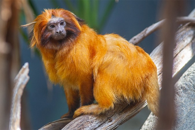
The morpho butterfly is a flying marvel. It flits through the rainforests of Central and South America. With wings that can span 20 centimeters (8 inches), it can be bigger than most human hands. Those wings shimmer with a dazzling blue hue. New data show these butterfly wings could one day become the basis of a new medical tool — one that might help doctors investigate the development and severity of some cancers.
Although this butterfly’s wings are blue, that hue is not due to any pigment. Instead, it comes from how light reflects and refracts off tiny microstructures atop those wings. The shimmering color of the wing depends on how light hits it. (Many materials, from rocks to plastic wrap, have this property, called structural color.)
Researchers at the University of California, San Diego (UCSD) have now shown how those microstructures can provide a new view on biological tissue.
The method they devised was simple. They placed a morpho wing on one microscope slide. Then they placed a thin slice of tissue on a second slide. After lining up the two, they turned on a special kind of light. This polarized light went through both the wing and the tissue beneath it. The light reflected back and passed through both again.

This process can reveal alterations in the structural fibers that make up certain tissues. These are called fibrotic changes. And they can help researchers diagnose or study many diseases.
The UCSD team described its new technique in the January 15 Advanced Materials. It can spot changes in fibrous tissue that may signal breast cancer — and whether that cancer is getting ready to spread.
It’s often hard to study fibrous tissues in a medical clinic, says Lisa Poulikakos. She’s a materials scientist at UCSD. “Usually,” she explains, “you need those expensive optical machines that cost around $200,000.” They’re not available to many doctors. Plus, she adds, looking for microstructural changes in tissue often requires first staining or dyeing it. But those processes change the tissue itself. The dyes also break down with time.
This new technique is a simpler and less expensive way to diagnose the fibrous nature of cancer tissue, she says. It requires only a microscope and one morpho wing. And the only thing that hits the tissue, she notes, is light.
Start with one pretty blue wing
The method came about because two scientists happened to be in the right place at the right time. Poulikakos was one of them. The other was Paula Kirya. This graduate student had just come to study at UCSD.
“Dr. Poulikakos was maybe the first person I met in San Diego,” says Kirya. “She let me join her lab very quickly.” In that lab, scientists study how light interacts with materials.
This team knew that fibrous tissue reflects light in unusual ways. They also knew that etching marks into a material could make it reflect different colors of light in different directions. They wanted to combine these two ideas to create a tool that would more accurately reveal how fibrous some tissue might have become.
When Kirya visited the lab, a student told her they were having trouble reflecting blue light. At once, something clicked in Kirya’s mind. She went to Poulikakos and said: “If this is what we’re trying to do, we could just look at the morpho butterfly and see these features.”
For two years before she came to UCSD, Kirya had been studying the blue morpho butterfly. She knew its wings’ deep hue doesn’t come from any natural dye or other chemical. Instead, it shimmers blue thanks to how tiny structures on its surface manipulate light.
Poulikakos was delighted at Kirya’s idea — and the new project was born.
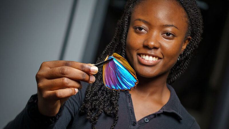
Kirya bought a single morpho wing for about $8 in a local store. “They gave us a discount,” she says. “Most insect collectors want pristine insects. We just wanted the wing.” Then she began experimenting with ways to combine samples of tumor tissue and this wing.
By rotating the tissue and wing together under a microscope, the scientists quickly found they could see the thick fibers associated with cancer.
Their method didn’t require any costly materials. Best of all? It needed no stain or dye. In fact, this is one major advantage of the new method, says Vance Williams. A materials scientist, Williams works at Simon Fraser University in Vancouver, Canada.
“The material itself doesn’t absorb light. And it’s not using dye that tends to fade over time,” he says. “It’s just all weird optical effects.”
Plus, Kirya notes, a single wing can be used again and again for years.
But this discovery was just the first step.
Polarizing the information
Polarized light is made up of light waves that all vibrate in the same plane. When the direction of this light lines up with tiny structures on the morpho’s wing, the light reflects brightly. The same thing happens for fibrous tissue, though less strongly.
The UCSD team found that shining polarized light through both materials now strengthened the light signal from those fibers.
“They’re using some clever optics,” Williams says, to make it “amplify a small signal.”
The light show put on by morpho butterfly wings shows up in other materials, too, he notes. (You can see much the same thing by looking through two polarizing lenses at stretched-out plastic wrap.) Williams designs materials with similar properties. They change color depending on how light strikes them.
Kirya may be young, says Poulikakos, but “she’s really a true scientist.” She took detailed measurements of the fibrous tissues and the reflected light. Then she worked with other researchers to develop a computer model to turn those measurements into meaningful information about how advanced a breast cancer was. That’s a gauge of how likely it is to spread and become lethal.
Kirya suspects that her new technique could be most useful in parts of the world where scientists don’t have access to expensive machines for diagnosing tissues. “I had been looking at butterflies for a long time,” she says. “It was a dream of mine to apply that knowledge to real life. To a real-life problem.” Now she has.
This is one in a series presenting news on technology and innovation, made possible with generous support from the Lemelson Foundation.